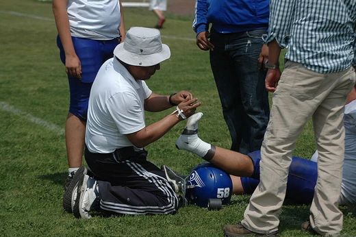
Cauliflower ear
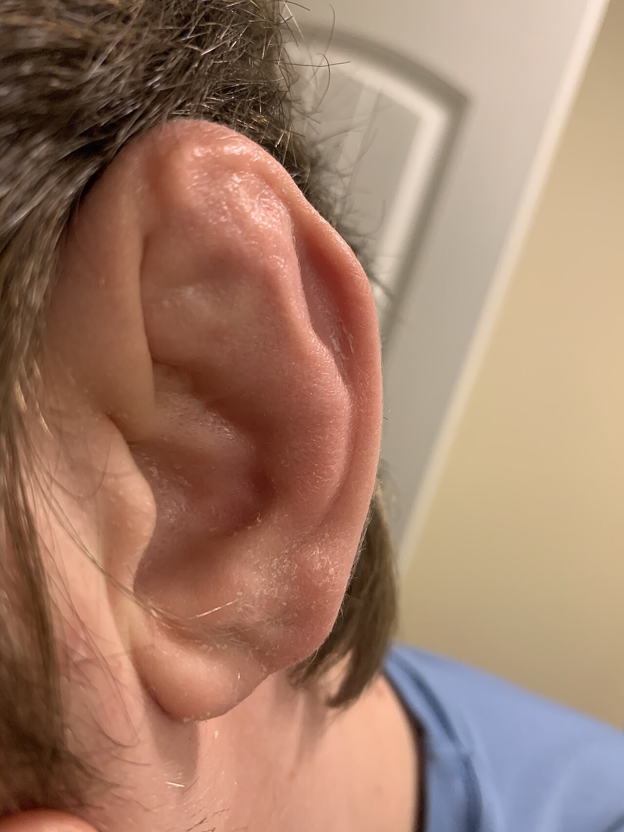
Irreversible ear deformity from untreated auricular hematoma.

Wikipedia / Wikimedia Commons
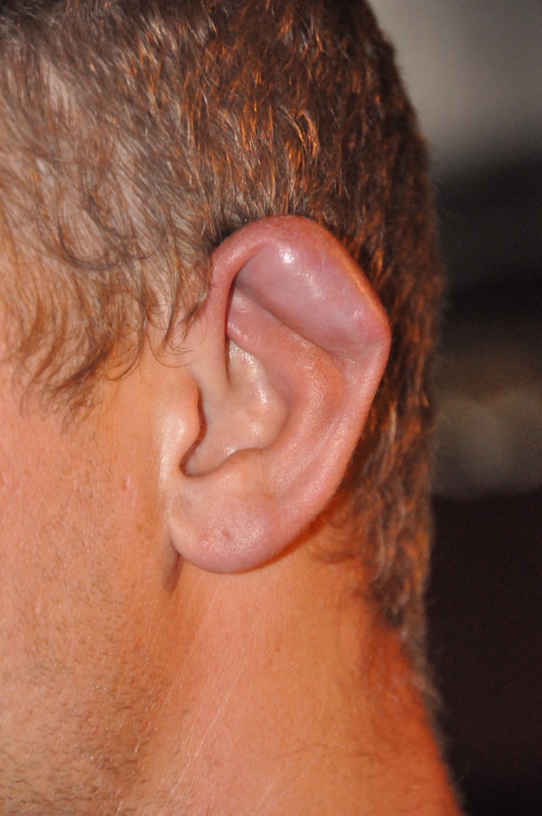
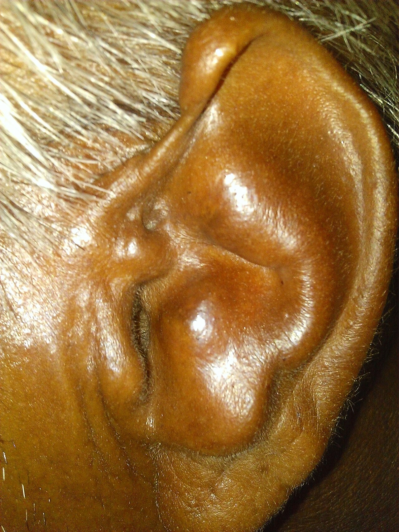
Cauliflower ear is an irreversible condition in which the external portion of the ear becomes permanently swollen and deformed, resembling a cauliflower. It occurs when blunt trauma to the ear causes a blood clot or fluid collection under the perichondrium, separating the cartilage from its nutrient supply and leading to cartilage death and fibrous tissue formation. The condition is common in contact sports such as wrestling, boxing, kickboxing, Brazilian jiu-jitsu, judo, sumo, mixed martial arts, rugby union, and rugby league.
- field
- Sports medicine, otolaryngology
- common_causes
- Blunt trauma to the ear, auricular hematoma
- sports_associated
- Wrestling, boxing, kickboxing, Brazilian jiu-jitsu, judo, sumo, mixed martial arts, rugby union, rugby league
- prevention
- Scrum cap, headgear, ear guard, specialty ear splint
- treatment
- Drainage of hematoma, pressure dressings, sutures, splints, antibiotics
Lore & Background
Cauliflower ear results from blunt trauma that causes an auricular hematoma, where blood accumulates between the perichondrium and cartilage. The hematoma obstructs blood flow, leading to cartilage necrosis and disorganized fibrosis. The condition is diagnosed clinically through history of injury and physical exam findings such as tenderness, bruising, and deformation of ear contours. A classification system by Yotsuyanagi et al. guides surgical decisions.
Reader's Guide
Cauliflower ear is significant primarily in contact sports and martial arts, where it is often considered a badge of courage or experience. Prompt evacuation of the hematoma within six hours is critical to prevent permanent deformity, though no single best treatment protocol has been identified. Drainage is proven superior to conservative treatment, but optimal bandaging or splinting after drainage requires further research. Without intervention, the ear can sustain serious damage including disruption of the ear canal and reduced blood flow. Cosmetic procedures may improve appearance. The condition was recorded in ancient Greece and noted in 19th-century Hong Kong opium dens from sleeping on hard wooden pillows.
Did You Know?
- Cauliflower ear can also result from nontraumatic inflammatory injury such as relapsing polychondritis, a rare rheumatologic disorder.
- The outer ear skin is tightly adherent to the perichondrium with almost no subcutaneous fat on the anterior surface, increasing hematoma risk.
- Auricular hematoma most often occurs in the space between the helix and antihelix (scapha) and can involve both anterior and posterior ear surfaces.
- Simple drainage becomes less useful after six hours from injury or with recurrent trauma, when open surgical treatment is more effective.
The Anatomy of a Permanent Mark
When a forceful blow or grinding shear force strikes the outer ear, the consequences unfold in a precise anatomical sequence. The skin covering the ear's cartilage is remarkably thin, with virtually no cushioning layer of subcutaneous fat on the front surface. This leaves the perichondrium—the vascular membrane that feeds the otherwise bloodless cartilage—exposed and vulnerable. When trauma ruptures the tiny vessels in that membrane, blood pools in the narrow gap between perichondrium and cartilage. That collection, called a hematoma, acts as a mechanical barrier, cutting off the nutrient supply the cartilage depends on. Starved of blood, the cartilage begins to die. Over time, disorganized fibrous tissue and irregular new cartilage fill the concave spaces of the pinna, kinking and deforming the helix, antihelix, concha, tragus, and antitragus. The result is a permanently swollen, lumpy outer ear that bears a striking resemblance to a head of cauliflower. The scapha, the groove between helix and antihelix, is the most frequent site, though the hematoma can spread into the triangular fossa, the concha, or even the area around the ear canal. In severe cases both the front and back surfaces are involved, which carries the highest risk of tissue death.
A Mark of the Mat
Cauliflower ear is inextricably linked to the world of combat and contact sports. The condition appears with particular frequency among wrestlers, boxers, kickboxers, and practitioners of Brazilian jiu-jitsu, judo, sumo, and mixed martial arts. It also shows up in full-contact team sports like rugby union and rugby league, where the constant head-to-head collisions create the same shear forces that damage the ear's delicate vascular supply. In these disciplines, the ear is not merely a casualty; for many athletes, the deformed pinna has become a visible badge of courage and accumulated experience, a physical testament to years of grappling and striking. In the immediate aftermath of injury, the affected ear typically feels tense, fluctuant, and tender, with a deep throbbing pain. Yet because the trauma that produces an auricular hematoma so often comes alongside more dramatic injuries—head lacerations, neck wounds, or other facial damage—the ear problem can easily slip past a clinician's notice unless specifically examined. The condition is irreversible once the cartilage has died and fibrous tissue has taken its place, making the prevention window narrow and the cultural weight of the deformity all the more significant in sports communities that wear it proudly.
Reading the Ear: Diagnosis and Staging
Because cauliflower ear and its precursor, the perichondral hematoma, are identified through clinical assessment rather than imaging, the diagnostic process relies heavily on the patient's story and a careful physical examination. A clinician will ask about participation in contact sports, the mechanism of the ear trauma, and whether similar episodes have occurred before. On examination, they look for tenderness over the affected area, visible bruising, and any distortion of the ear's normal contours. The combination of history and physical findings is sufficient to confirm the diagnosis and guide the choice of treatment. To bring more structure to clinical decision-making, Yotsuyanagi and colleagues developed a classification system specifically for auricular hematomas. Their framework helps providers determine at what point surgical intervention becomes necessary and offers a roadmap for selecting the most appropriate surgical approach for a given severity. This is particularly important because the condition can be easily missed when the patient presents with more conspicuous injuries elsewhere on the head or neck, and because the window for effective intervention is limited. Without directed attention to the ear specifically, a treatable hematoma can progress silently into permanent deformity.
Draining, Splinting, and Wearing the Mark
Treatment of an acute auricular hematoma centers on one principle: getting the pooled blood out before the cartilage loses its blood supply permanently. Research consistently shows that draining the hematoma outperforms simply watching and waiting, though no single protocol has emerged as the definitive standard. In the acute phase, clinicians may use needle aspiration, make a small incision to let the blood escape, apply continuous suction devices, or pack a wick into the wound. Once the fluid is evacuated, the priority shifts to preventing re-accumulation. A toolkit of compression methods has been developed: direct-pressure dressings, mattress sutures pinning skin back to cartilage, small buttons threaded onto sutures, thermoplastic splints molded to the ear, sutured cotton balls, and absorbable mattress sutures. Simple drainage loses effectiveness roughly six hours after injury, and with recurrent trauma, open surgical repair becomes the more reliable route to restoring the ear's shape. On the prevention side, rugby players wear scrum caps, while wrestlers and martial artists use headgear or dedicated ear guards. A custom ear splint can keep the damaged area compressed to block fluid buildup. For some competitors, however, the resulting deformity is worn as a badge of honor.
Gallery

Frequently Asked Questions
What is cauliflower ear?
Cauliflower ear is a permanent deformity of the outer ear in which the cartilage swells and hardens into a lumpy, cauliflower-like shape. It occurs when the ear's cartilage loses its nutrient supply after trauma and is gradually replaced by fibrous scar tissue.
How does cauliflower ear develop?
A blunt hit to the ear creates a blood clot or fluid pocket beneath the perichondrium, pulling the cartilage away from the tissue that feeds it. Once the cartilage dies, the body fills the gap with scar tissue, locking in the deformed shape permanently.
Which sports put athletes at the highest risk for cauliflower ear?
The condition is most commonly seen in grappling and striking disciplines such as wrestling, Brazilian jiu-jitsu, judo, sumo, boxing, kickboxing, and mixed martial arts, as well as rugby union and rugby league where repeated head contact is part of the game.
Can cauliflower ear be reversed or surgically corrected?
Once the cartilage has died and fibrous tissue has formed, the deformity is irreversible and no treatment can restore the ear to its original shape. Early medical intervention—draining the hematoma, applying pressure dressings, sutures, splints, and sometimes antibiotics—can stop the condition from setting in the first place.
What can athletes do to prevent cauliflower ear?
Wearing protective gear such as a scrum cap, headgear, a dedicated ear guard, or a specialty ear splint during training and competition significantly reduces the chance of the initial impact that triggers the condition.
More in Sports injuries 1-24
Spotted an error? Know more?
This is a living reference — every entry is fact-audited, and reader corrections feed straight into our audit queue. Suggest an edit · See this site's audit record